

Izmir: 8cm Brain Tumor Removed With Hybrid Method, Heralded as Medical Success
Translated from Turkish, summarized and contextualized by DistantNews.
At a glance
News
Sources not specified
Outcome reported
- Doctors successfully removed an 8-centimeter brain tumor from a patient in Izmir using a hybrid surgical method.
- The patient, Songül Bacak, experienced symptoms like dizziness and headaches for two years before diagnosis.
- The procedure involved blocking the tumor's blood vessels before surgical removal, minimizing bleeding risks.
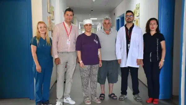
In a medical feat that may enter the literature, doctors in Izmir have successfully removed an 8-centimeter brain tumor using a hybrid technique. Songül Bacak, a resident of Uşak, had been experiencing symptoms for two years, including brief moments of fainting, visual disturbances, headaches, and nausea, which she did not initially recognize as seizures.
Her condition worsened, prompting her to seek medical attention. Diagnostic tests revealed a significant tumor, approximately 8 centimeters in size, in her brain. She was transferred to SBÜ İzmir Tepecik Training and Research Hospital, where she underwent treatment. A multidisciplinary team, comprising specialists from Interventional Radiology and Neurosurgery, performed a joint operation on July 7 to remove the tumor.
I had headaches and nausea for two years, and I saw fleeting visions. I only learned here that these were seizures. My doctor explained all the risks from the beginning, but he gave me such great confidence that I went into surgery without fear. When I came out of surgery and opened my eyes, I felt like I was reborn into the world. I am so happy.
Bacak shared her experience, stating she felt no fear upon learning about the tumor due to her complete trust in her doctors. "I had headaches and nausea for two years, and I saw fleeting visions. I only learned here that these were seizures. My doctor explained all the risks from the beginning, but he gave me such great confidence that I went into surgery without fear. When I came out of surgery and opened my eyes, I felt like I was reborn into the world. I am so happy," she said.
When we heard she was having brain surgery, of course, we were very scared. We signed the consent form, accepting everything, including the risk of death. We trusted our doctors. Thank God, we got through that ordeal, and we are very pleased now.
Her husband, Ferhat Bacak, 51, expressed immense relief at his wife's recovery. "When we heard she was having brain surgery, of course, we were very scared. We signed the consent form, accepting everything, including the risk of death. We trusted our doctors. Thank God, we got through that ordeal, and we are very pleased now," he stated.
Dr. Serhat Akış, an Interventional Radiology Specialist, explained the innovative approach. "Our patient had a very large tumor, about 8 centimeters, at the head. Operating on lesions of this size carries serious bleeding risks. Therefore, before the surgery, we mapped the brain vessels using the brain angiography method in interventional radiology. Using our advanced angiography devices, we reached only the vessels feeding the tumor without harming healthy tissue and blocked these vessels with special closure devices. Thus, we prepared the tumor for surgery, so to speak. This method reduces bleeding during surgery and also makes it easier for the surgeon to distinguish between live and dead tissue, offering a much safer and more comfortable surgery," Dr. Akış noted.
Our patient had a very large tumor, about 8 centimeters, at the head. Operating on lesions of this size carries serious bleeding risks. Therefore, before the surgery, we mapped the brain vessels using the brain angiography method in interventional radiology. Using our advanced angiography devices, we reached only the vessels feeding the tumor without harming healthy tissue and blocked these vessels with special closure devices. Thus, we prepared the tumor for surgery, so to speak.
Assoc. Prof. Dr. Alper Tabanlı, Head of the Neurosurgery Training Clinic, confirmed that the operation on July 7, lasting approximately 6 hours, was successfully completed. "When our patient arrived, we saw a quite large lesion of 8-9 centimeters in the tests," he added.
This method reduces bleeding during surgery and also makes it easier for the surgeon to distinguish between live and dead tissue, offering a much safer and more comfortable surgery.
DistantNews Editorial
Originally published by Cumhuriyet in Turkish. Translated, summarized, and contextualized by our editorial team with added local perspective. Read our editorial standards.
More Stories


From Our Blog

What Gets Lost (and Found) When News Is Translated

The Critical Role of Diaspora Media in Global News

What Travellers Should Know About Countries with Restricted Press

A Digital Nomad's Guide to Following Local News

How the Same Story Looks Different in Different Countries
